Case Report
Austin J Surg. 2022; 9(2): 1289.
Management of an Uncommun Giant Ovarian Hydatid Cyst
Dhaou AB¹*, Karmous N² and Kerkeni A³
¹Visceral and Digestive surgeon, Unit “A” of General Surgery, Tunis El Manar University, Tunisia
²Department “B” of Obstetrics and Gynecology, University Tunis El Manar, Tunisia
²Visceral and Digestive surgeon, Unit “A” of General Surgery, Tunis El Manar University, Tunisia
*Corresponding author: Anis Ben Dhaou, Visceral and Digestive surgeon, Unit “A” of General Surgery, Charles Nicolle Hospital, Tunis, Faculty of Medicine of Tunis, Tunis El Manar University, Tunisia
Received: August 02, 2022; Accepted: August 26, 2022; Published: September 02, 2022
Case Report
We report the case of a 52-year-old patient. She was overweight and thought at first she was gaining weight.
She presented to our department with a gradually increasing huge abdomen after 11 months of clinical evolution. She was not febrile and had normal vital signs on general assessment. No icterus or edema was present. On examination of the abdomen, there was widespread distension and a dull tone on percussion. The liver and spleen could not be felt.
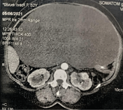
A large abdominal echogenic tumor filled the whole abdomen and pelvic cavity, according to the CT scan (Figure 1). Despite their rarity, peritoneal cystic mesothelioma and abdominal cystic lymphangioma should be suspected in the presence of such a pelvic mass. The huge cyst was confirmed on an abdomino-pelvic Magnetic Resonance Imaging (MRI) and measured 65 x 52 cm, supero-laterally displacing the liver and spleen. The kidneys were located in the back, and the bowel loops were located in the front. The ovaries were not visible. There was fluid in the peritoneal cavity (Figures 2 and 3). Tumor markers (CEA, a-fetoprotein and CA-125) were within normal limits. The hydatid cyst serology was positive.

Figure 1: CT scan of the giant cyst.

Figure 2: MRI coronal section of the giant cyst.

Figure 3: MRI axial section of the giant cyst.
The rural origin of our patient, especially in an endemic zoonotic area (Tunisia), as well as the morphological examination of the mass (a cystic mass with its own wall), led us to evoke the diagnosis of an intrapritoneal primary hydatid pelvic cyst even though it is extremely rare.
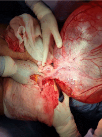
The patient underwent explorative laparotomy for diagnostic and therapeutic purposes. The mass was so large that it could not be removed without a large abdominal incision. The mass was extracted without perforation to avoid any hypothetical hydatid infestation of the peritoneal cavity (Figure 4). The right ovary was included in the mass and the right fallopian tube was adherent to the surface of the cyst. Complete excision of the cyst with left salpingo-oophorectomy was performed. The specimen weighed 20 kg (Figure 5 and 6). It had no visible exovesiculation because there were no sources of aggression such as in the case of the hydatid hepatic cyst where the aggression is due to the bile.

Figure 4: Right ovary and fallopian included in the hydatid giant cyst.

Figure 5 and 6: The hydatid giant cyst extracted.
Histopathological report revealed hydatid cyst of the ovary. The patient was discharged on the sixth day following surgery after an uneventful postoperative period. The patient was given albendazole for preventive purposes.