
Research Article
Austin J Radiol. 2024; 11(1): 1225.
Dosimetric Comparison of VMAT Plans by Varying Number of Arcs Forhead and Neck Cancer Patients
Harijith KR1; Jitendra Nigam2; Silambarasan NS3; Navitha S3*; Piyush Kumar4
1Intern Medical Physicist, SRMS Institute of Medical Science, Department of Radiation Oncology, Shri Ram Murti Smarak Institute of Medical Sciences, Bareilly
2Associate Professor cum Medical Physicist, Department of Radiation Oncology, Shri Ram Murti Smarak Institute of Medical Sciences, Bareilly
3Assistant Professor cum Medical Physicist, Department of Radiation Oncology, Shri Ram Murti Smarak Institute of Medical Sciences, Bareilly
4Professor and Head, Department of Radiation Oncology, Shri Ram Murti Smarak Institute of Medical Sciences, Bareilly
*Corresponding author: Navitha Silambarasan Department of Radiation Oncology, Shri Ram Murti Smarak Institute of Medical Sciences, Bareilly. Tel: +91 9677760497 Email: navitha.selvi@gmail.com
Received: December 29, 2023 Accepted: February 03, 2024 Published: February 10, 2024
Abstract
Aim: This study is intended to evaluate the plan quality using Volumetric Modulated Arc Therapy by increasing number of arcs.
Materials and Methods: Twenty patients diagnosed with various head and neck cancers were selected for this study. The patients were treated using Varian Truebeam linear accelerator with VMAT dual arc. These patients were replanned using VMAT plans employing one, three, and four arcs through the Varian Eclipse
13.6 treatment planning system. The target delineation and Organ-At-Risk (OAR) contours were done by the radiation oncologist as per RTOG guidelines. With a grid size of 2.5mm, the dose distribution was determined using the Anisotropic Analytic Algorithm (AAA) and the constraints were kept constant across all plans. All plans were optimized using Progressive Resolution Optimizer. Plan quality was assessed using the Conformity Index, Homogeneity Index, and by evaluating the Planning Target Volume (PTV) coverage, D2, D98, D50 and the dose to the OAR using Dose-Volume Histogram (DVH). Monitor units were also evaluated. Two-way evaluation of plan was done; one is by visually checking the isodose coverage and other using DVH. Statistical data were analyzed using a student’s t-test.
Result: Clinically acceptable target coverage was achieved in all plans. The four-arc plan yielded a significant Conformity Index (P<0.05) and Homogeneity Index (P<0.05) compared to other plans. The four- arc plans resulted in a significant dose reduction for PRV (Planning Organ at Risk Volume) Spine, lips and parotid. There was no significant difference between the dose to PRV Brainstem and cochlea. A lower Monitor Unit (MU) per 2.0 Gy per fraction was achieved using 1 Arc (436 MU), 2 Arcs (505 MU), 3Arcs (464 MU), and 4 Arcs (486 MU). Hence, reduced treatment time was observed in the one-arc VMATplan.
Conclusion: The conformity index and Organ-At-Risk (OAR) dose improved with an increase in the number of arcs from two to four. Therefore, utilizing a higher number of arcs in VMAT plans can enhance plan efficiency and reduce the dose to OAR. The results suggest that a 4-arc VMAT plan may serve as an alternative to the 2-arc plan, offering improved plan quality and reduced OAR dose in head and neck cancer. As the number of arcs increases the integral dose also increases, so only two arc is necessary for treating young patient to avoid secondary malignancy.
Keyword: VMAT; Arcs; Head and Neck cancer; Conformity Index; Homogeneity Index
Abbreviations: AAA: Analytic Anisotropic Algorithm; CI: Conformity Index; HI: Homogeneity Index; DVH: Dose Volume Histogram; VMAT: Volumetric Modulated Arc Therapy; PTV: Planning Target Volume; CT: Computer Tomography; IMRT: Intensity Modulated Arc Therapy; CTV: Clinical Target Volume; MV: Megavoltage; OAR: Organ at Risk Volume; PRV: Planning Organ at Risk Volume; TPS: Treatment Planning System; GTV: Gross tumor Volume; CECT: Contrast enhanced Computer Tomography; MLC: Multi-Leaf Collimator; PRO: Progressive Resolution Optimizer; DVH: Dose Volume Histogram; RTOG: Radiation Therapy Oncology Group; EBRT: External Beam Radiation Therapy; DICOM: Digital Imaging and Communication in Medicine
Introduction
Head and neck cancer is the seventh most common type of cancer worldwide and comprise of diverse group of tumors affecting the upper aero digestive tract. Head and Neck cancer include cancer in larynx, lips, throat, nose, salivary gland [1]. The type of treatment recommended depends on the location, site and type of cancer.
The head and neck cancer were selected because it’s a challenging scenario for treatment planning. Inappropirate dosing may result in either recurrence of disease or severe toxicity, thus it’s important to investigate the novel radiation delivery technique improves the dose coverage to target volume and minimal dose to OAR. Treatment options for patients with head and cancer include fractionated External Beam Radiotherapy (EBRT) or surgery (combined with EBRT), either with or without chemotherapy [1].
From all modalities used for treating cancer, radiation therapy seems to be a significant feature for effective treatment for Head and Neck cancer. With the introduction of modern radiation therapy technique such as Intensity Modulation Radiation Therapy (IMRT) and Volumetric Modulated Arc Therapy (VMAT), radiation side effects during treatment are reduced. Therefore, IMRT and VMAT are chosen for radiation therapy for Head and Neck cancer.
The term Intensity-Modulated Radiation Therapy (IMRT) refers to a radiation therapy technique in which a non-uniform fluence is delivered to the patient from any given position of the treatment beam to optimize the composite dose distribution. The treatment criteria for plan optimization are specified by the planner and the optimal fluence profiles for a given set of beam directions are determined through inverse planning [2].
IMRT is highly effective in treating target structure with irregular contour, while reducing dose to healthy tissues. VMAT is a novel form of IMRT in which radiation treatment is delivered during gantry rotation with dynamic multi leaf collimator motion, variable dose rate and gantry speed modulation [2]. The main advantage of VMAT is less treatment time and reduced MU over conventional IMRT. Many studies have shown that VMAT can produce dosimetrically equivalent plans to IMRT for centrally located tumor such as prostate cancer, cervical cancer and head and neck cancer [3-4].
The beam on time for VMAT may be less than 3 mins. Since the Rapid arc optimization tries to maximize the gantry speed and dynamic MLC motion, we hypothesized that using more than two arcs, which would allow for a longer delivery time and more opportunity for modulation, might translate into further gains in plan quality. In search for better VMAT plans there are many parameters that the planar can modify, including the number of arcs.
Many considerations have been kept forward to enhance the plan quality in VMAT plans, including the number of beam arcs, may be chanced in quest for better volumetric modulated arc therapy plans. The present study aimed to compare the Dosimetric parameters of VMAT plans with varying number of arcs for head and neck cancer patients.
Materials and Methods
Patient Selection and Simulation
CT dataset was identified for twenty patients (15 males and 5 females) of head and neck cancers were selected for this study. All patients were immobilized with Klarity five push pin head and neck thermoplastic cast in the Head and Neck base frame. Patients were positioned in supine position with their arms alongside their body. All the CT scans were taken using contrast (CECT) which is use differentiate tumor volume from others. All of the CT dataset were acquired using a Simens Somato Scope CT (32 Slice) scanner. The CT image was taken at 3 mm slice thickness. The CT images were taken from supra orbital to trachea bifurcation for Head and Neck cancer. The data were transferred to the Eclipse 13.6 treatment planning system using DICOM format.
Delineation of Target Volume and Organ at Risk
Radiation oncologist contoured the target volume as per RTOG guidelines. Target structure such as Gross Tumor Volume (GTV), Clinical Target Volume (CTV), planning target volume and the Organ at Risk volume (OAR) were contoured. To ensure that the recommended dosage is administered to CTV and reduce the risk of treatment failure due to variability in position setup and movements of the organ during actual treatment, the planning target volume were obtained by expanding CTV, 5 mm in all directions expect in the direction of skin. The Organ at risk volume such as the spinal cord, Brainstem, parotid, Mandible, lips, cochlea were contoured by the radiation oncologist. An extra 5 mm margin was added to spinal cord and 3 mm added to brainstem as the planning organ at risk volume.
Treatment Planning
VMAT plans were created for all 20 patients using 6 MV X-ray photon beam energy from a Varian Truebeam linear accelerator which was equipped with an MLC with 120 leaves (of thickness 1cm for outer 10 pairs and 0.5 cm for inner 40 pair leaves). Four plans were created for each patient including single arc, dual arc, three arc and four arc plans optimized using Progressive Resolution Optimizer (PRO), eclipse treatment planning system version 13.6. (Varian medical system). The pictorial Representation of beam orientation of VMAT plans using single Arc, dual Arc, three Arc andfour Arc plans is shown in Figure 1. All the plans were optimized at a maximum DR of 600MU/min. The isocenter was taken as the center of the PTV. With a grid size of 2.5mm, the dose distribution was determined using the Anisotropic Analytic Algorithm (AAA) and the constraints were kept constant across all plans. The planning objective for PTV was at least 95% of the PTV volume receives 95% of the prescribed dose. During optimization the PTV was given the max priority and the Normal tissue objective was selected as, distance from target border as 3mm, start dose at 105% and end dose at 60%, dose fall off 3 mm. The normal tissue objective was given a priority of 100.
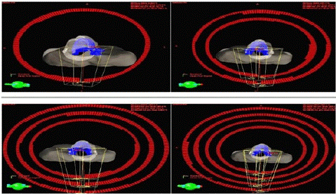
Figure 1: Representation of beam orientation of VMAT plans using single Arc dual Arc, three Arc andfour Arc plans.

Figure 2: The reference isodose distribution on transverse view for one patient planned by single arc, dual arc, three arcs and four arcs VMAT plans.
Plan Evaluation
The plan quality indices of all the VMAT treatment plans were analyzed using Dose Volume Histogram (DVH), which represent the whole dose volume information in a two-dimensional single curve. Coverage of PTV volume and mean and max dose to OAR and conformity and homogeneity index were analyzed for every plans. The OAR dose was evaluated based on RTOG. The ratio of volume of PTV covering reference isodose of prescribed dose to the product of volume PTV and volume of reference isodose was used to calculate conformity index [5]. the conformity index describes the degree to which the prescribed isodose volume conforms to the shape and size of the target volume.
The ratio of difference between to dose received by 2% volume of PTV and 98% of PTV to the dose received by 50% of PTV was used to calculate the Homogeneity index. A homogenous plan is defined as an HI value close to zero. (6). The homogeneity index describes the uniformity of dose within a target volume and is directly calculated from the statics of Dose Volume Histogram.
The PTV structures were examined for D98%, D2%, and D50%. The doses to the OAR were evaluated using DVH. The max dose to PRV spine and brainstem and the mean dose to parotid and lips and cochlea were evaluated. The monitor units for all the plans were evaluated. For Statistical evaluation of the plan, student’s t-test was used to see which of the evaluated factors improved significantly by using a greater number of arcs, with a p value<0.05.
Result
The target dose homogeneity, conformity index, monitor unit, treatment delivery time and OAR sparing all improved as the number of arcs was increased.
Target Coverage, Conformity and Dose Homogeneity
All the VMAT plans were clinically acceptable. Acceptable target coverage (95% of prescribed isodose covering 95% of the target volume) was achieved in all the plans but statistically the four arc VMAT plans generated significantly better conformity and homogeneity. The conformity and homogeneity of the VMAT plans improved as a greater number of arcs were used. The color wash of the reference isodose distribution is shown in Fig 2. The Conformity index and Homogeneity index is significant (P<0.05) in four arc plans compared to other plans. There was no significant difference between two arc and three arc plans. The conformity and homogeneity index improved as the number of arc increases.
One arc plan shows less homogenous dose distribution compared to other plans. The MU was less in one arc plan. The D2%, D98% and D50% of PTV for one Arc plan is significant compared to other plans. There was no significant difference between two arcs, three arc plans and four arc VMAT plan. TheD2%, D98% and D50% of PTV for all plans is showed in Figure 3.
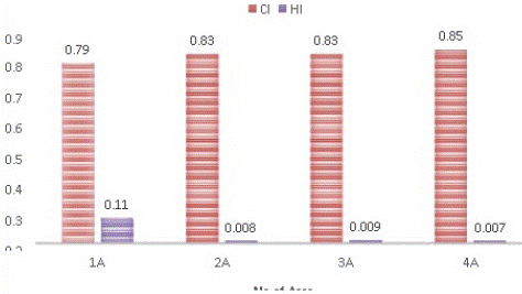
Figure 3: Histogram showing the dose Conformity Index and Homogeneity Index of one Arc, dual Arc, threeArc and four Arc plan.
The one arc VMAT plan produced less conformity than other plans and dose distribution is not homogenous compared to other VMAT plan. All the VMAT plans achieved acceptable target coverage, and the hotspot was more in one arc VMAT plan compared to other plans.
Dose to Organ at Risk
The quantitative comparison of OAR dose among four different are summarized in Table 2. Dose constrains for all healthy tissue structure were all within the tolerance limits. Increasing the number of arcs, lead to an improving dose sparing to healthy tissues.
![]()
    Parameters
    Number of Arc’s
    P VALUE


    CI
    0.79±0.05
    0.83±0.04
    0.83±0.05
    0.85±0.05
    0
    0
    0.0002
    0.098
    0.0025
    0.0503


    HI
    0.11±0.02
    0.08±0.02
    0.09±0.01
    0.07±0.02
    0
    0
    0
    0.0328
    0.0004
    0.042


    MU
    436.5±84
    505.6±109
    464.2±86
    486.45±91
    0
    0.0005
    0
    0.0025
    0
    0.0857


    D 2% (Gy)
    63.5±15
    65.24 ±7
    65.33±7
    65.24±7
    0.2462
    0.236
    0.2472
    0.1999
    0.0001
    0.4828


    D98%(Gy)
    56.55±13
    59.79±6
    59.78±6
    59.93±6
    0.0792
    0.0787
    0.0699
    0.048
    0.0038
    0.0236


    D50%(Gy)
    61.06±14
    63.45±7
    63.46±7
    63.45±7
    0.1648
    0.1643
    0.1648
    0.4589
    0.3255
    0.4888


    D95%(Gy)
    60.62±5.5
    60.66±5.5
    60.63±5.5
    60.63±5.5
    0.362
    0.1765
    0.1632
    0.0664
    0.2862
    0.1012


    (P1:    1ARC VS 2ARC, P2: 1ARC VS 3ARC, P3: 1ARC VS 4ARC, P4:2ARC VS 3ARC, P5: 3ARC    VS 4ARC, P6: 2ARC VS 4ARC, P Value<0.05-Significant)
Table 1: Comparison of conformity, homogeneity and MU of all the VMAT plans and p value.
![]()
OAR
Number of Arcs
PP Value
1A
2A
3A
4A
P1
P2
P3
P4
P5
P6
PRV BRAINSTEM
(Dmax)36.05±12.6
35.01±12.4
35.53±12.5
35.17±12.4
0.077
0.1782
0.1105
0.108
0.111
0.3483
PRV SPINE
(Dmax)45.24±4.3
43.26±4.6
43.67±4.5
42.81±4.2
0.0000
0.0000
0.0000
0.041
0.0409
0.046
RT PAROTID
(Dmean)33.44±12.3
32.61±12
33.14±12.4
32.83±12.4
0.0034
0.2155
0.0469
0.019
0.0475
0.2988
LT PAROTID
(Dmean)31.29±9.2
30.04±8.2
30.07±8.7
29.58±8.2
0.014
0.1785
0.0005
0.166
0.1606
0.0539
LIPS (Dmean)
27.19±9.9
26.79±9.4
26.84±9.9
26.13±9.5
0.1452
0.1446
0.0126
0.44
0.0083
0.0458
LTCOCHLEA
(Dmean)16.14±12.7
14.82±11.6
16.69±12.8
15.79±11.8
0.1662
0.1392
0.3736
0.112
0.1254
0.2926
RTCOCHLEA
(Dmean)14.78±12.1
16.48±11.9
15.64±11.9
16.07±12.5
0.1663
0.0489
0.0748
0.31
0.3081
0.4142
(P1: 1ARC VS 2ARC, P2: 1ARC VS 3ARC, P3: 1ARC VS 4ARC, P4:2ARC VS 3ARC, P5: 3ARC VS 4ARC, P6: 2ARC VS 4ARC, P Value<0.05-Significant)
Table 2: Dosimetric Results of Organ at Risk (OAR).
For spinal cord all four plans reached the planning objective of Dmax <50 Gy. The Dmax of PRV spinal cord were significant in four arc plans compared to other plans, the Dmax values of spine were similar in dual arc and three arc. The dose to spine was more in one arc plan.
There was no significant difference between the dose to Brainstem in all the plans. The four arc VMAT plan generated a significantly (<0.05) lower dose to lips and parotid and no significant difference were obtained dose to cochlea. The DVH for the OAR of one patient is shown in Figure 4.
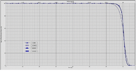
Figure 4: Comparison of dose volume histogram for PTV for four different plans.

Figure 5: Histogram showing the dose the OAR for one Arc, dual Arc, three Arc and four Arc plan.
Discussion
A study conducted by Li-Rong Zhao et al [11] on Comparison of plan optimization for single and dual volumetric modulated arc therapy verses intensity modulated radiation therapy during post- mastectomy regional irradiation. The results stated that plan quality and delivery efficiency made VMAT a reasonable option for post mastectomy regional irradiation, while 2ARC VMAT resulted in an improved HI and CI compared to 1ARC plan. The results of this study show that using 4ARC VMAT plans resulted in an improved HI and CI compared to other plans. As the number of ARC are increased the plan quality also increased in Head and Neck cancer.
A study conducted by Wu-Zhe Zhang et al [9] on Volumetric Modulated Arc Therapy vs c-IMRT for treatment of Upper Thoracic Esophageal cancer. The results stated that the dose conformity of PTV and OAR were improved when the arc number of the VMAT plan was increased from one to two for Esophagus cancer. The treatment time increased as more than 2ARC were used. The results of this study also show that the dose conformity of PTV and OAR improved when the number of arcs in VMAT plan increased from two to four in Head and Neck cancer. Increasing the number of arcs also increased the treatment time.
A study conducted by Sivakumar Radhakrishnan et al [12] on Dosimetric comparison between Single and Dual Arc-Volumetric Modulated Arc Radiotherapy and Intensity Modulated Radiotherapy for Nasopharyngeal Carcinoma using Simultaneous Integrated Boost Technique. The results stated that increasing the number of arcs provide additional flexibility in shaping the dose distribution. DA-VMAT showed better target coverage and achieved better sparing of OARs among the other plans. The results of this study also show that increasing the number of arcs provided additional flexibility in shaping the dose distribution as the plan quality increases when the number of arcs increases from two to four arcs.
A study conducted by D Kumar et al [10] on A comparative study of the dosimetric impact on IMRT planning with VMAT plans using a varying number of arcs in prostate cancer. The results suggested that the plan efficiency improved when higher number of arcs were used in VMAT plan for Prostate cancer. The four arc VMAT plans seemed to be a good compromise between faster delivery and high treatment plan efficiency in Prostate cancer. The results of this study also show plan efficiency improved when higher number of arcs were used in VMAT plan for Head and Neck cancer. The four arc VMAT plan resulted in increasing plan efficiency in Head and Neck cancer.
A study by Anne Richter et al [13] on Impact of beam configuration on VMAT plan quality for Pinnacle3Auto-Planning for head and neck case. The results showed that that double and full arcs are superior to single and partial arcs in terms of organs at risk sparing even for unilateral target volumes. The results of this study shows that four arc VMAT plan provided better dose sparing than other VMAT plan.
A study by Hani Ashamalla et al [14] on Comparison study of intensity modulated arc therapy using single or multiple arcs to intensity modulated radiation therapy for high-risk prostate cancer. The results showed VMAT appears to improve treatment efficiency, dosimetry, and conformity for patients with high- risk prostate cancer when compared to IMRT. The results of this study showed VMAT plans produced better improve treatment efficiency and conformity for patient when increasing the number of arcs in Head and Neck cancer.
Integral dose is an important consideration when developing plans with modulated arcs. Integral dose is the volume of dose deposited in the patient and is equal to the mean dose times the volume. The integral dose is more in IMRT plan than is conventional radiotherapy. It’s often stated that large number of beamlets and mu used in IMRT may lead to an increase in Integral dose, which may cause an increased risk of secondary malignancy [15].
In VMAT we have dose going through the body at 360 degrees. So integral dose becomes more important factor in areas with critical structure. As the number of arcs increase the integral dose increases and the risk of secondary malignancy also increases. Hene four arc VMAT plan may have more integral dose than other plans. For younger patients we don’t want any low dose volume and reduced Integral dose to reduce secondary malignancy is needed, so two arc VMAT plans in enough for young patients. Similarly, for elder patient in order to obtain best plan quality, four arc VMAT plan can be used. Our results showed that the number of MU required using one Arc (436 MU), two Arcs (505 MU), three Arcs (464 MU), four Arcs (486 MU). An increase in the number of MUs may increase undesirable irradiation of normal tissues via the scattered dose, leading to an elevated risk of secondary cancers after treatment. Hence the resulting increase in plan quality comes at an expense of increased delivery time.
Conclusion
The Conformity index and Homogeneity improved and dose to OAR reduced as the number of Arcs increases. Utilizing a higher number of Arcs in VMAT plan can enhance the plan quality and reduce dose to OAR. The resulting increase in plan quality comes at an expense of increased delivery time. Increasing the number of Arcs provided an additional flexibility in shaping dose distribution. Four arc VMAT plans can be used for treatments where the patient load is less as the treatment time is longer than other VMAT plan. As the number of arcs increases the integral dose also increase, hence two arc plan is used for treating young patients. Advantages of using multiple arcs and noncoplanar beams are now being fully explores.
Author Statements
Competing Interest
The author has declared that no competent interest exists.
References
- Mody MD, Rocco JW, Yom SS, Haddad RI, Saba NF. Head and Neck cancer. Lancet. 2021; 398: 2289-99.
- Wolff D, Stieler F, Welzel G, Lorenz F, Abo-Madyan Y, Mai S, et al. Volumetric modulated arc therapy (VMAT) vs. serial tomotherapy, step-and-shoot IMRT and 3D-conformal RT for treatment of prostate cancer. Radiother Oncol. 5th ed. 2009; 93: 226-33.
- Wagner D, Christiansen H, Wolff H, Vorwerk H. Radiotherapy of malignant gliomas: comparison of volumetric single arc technique (Rapid Arc), dynamic intensity-modulated technique and 3D conformal technique. Radiother Oncol. 2009; 93: 593-6.
- Ian P. A simple scoring ratio to index the conformity of radio surgical treatment plans. J Neurol Surg. 2000; 93: 219-22.
- The International Commission on Radiation Units and Measurements. Prescribing, recording and reporting photon-beam IMRT. J ICRU. 2010; 10: NP.2-NP.
- Cozzi L, Dinshaw KA, Shrivastava SK, Mahantshetty U, Engineer R, Deshpande DD, et al. A treatment planning study comparing volumetric arc modulation with RapidArc and fixed field IMRT for cervix uteri radiotherapy. Radiother Oncol. 2008; 89: 180-91.
- Guckenberger M, Richter A, Krieger T, Wilbert J, Baier K, Flentje M. Is a single arc sufficient in volumetric-modulated arc therapy (VMAT) for complex-shaped target volumes? Radiother Oncol. 2009; 93: 259-65.
- Zhang WZ, Zhai TT, Lu JY, Chen JZ, Chen ZJ, Li DR, et al. Volumetric Modulated Arc Therapy vs. c-IMRT for the Treatment of Upper Thoracic Esophageal Cancer. PLOS ONE. 2015; 10: e0121385.
- Kumar D, Pradhan A, Singh LM. A comparative study of the dosimetric impact on IMRT planning with VMAT plans using a varying number of arcs in prostate cancer. J Phys Conf Ser. 2021; 2070: 012011.
- Zhao LR, Zhou YB, Sun JG. Comparison of plan optimization for single and dual volumetric-modulated arc therapy versus intensity-modulated radiation therapy during post-mastectomy regional irradiation. Oncol Lett. 2016; 11: 3389-94.
- Radhakrishnan S, Chandrasekaran A, Sarma Y, Balakrishnan S, Nandigam J. Dosimetric comparison between single and dual arc-volumetric modulated arc radiotherapy and intensity modulated radiotherapy for nasopharyngeal carcinoma using a simultaneous integrated boost technique. Asian Pac J Cancer Prev. 2017; 18: 1395-402.
- Richter A, Exner F, Bratengeier K, Polat B, Flentje M, Weick S. Impact of beam configuration on VMAT plan quality for Pinnacle3Auto- Planning for head and neck case. Radiat Oncol. 2019; 14: 12.
- Ashamalla H, Tejwani A, Parameritis I, Swamy U, Luo PC, Guirguis A, et al. Comparison study of intensity modulated arc therapy using single or multiple arcs to intensity modulated radiation therapy for high-risk prostate cancer. Radiat Oncol J. 2013; 31: 104-10.
- Massat MB, et al. VMAT: the next generation of IMRT. Applied radiation oncology.