
Clinical Image
Ann Hematol Onco. 2024; 11(2): 1449.
Blood Smear as A Clue to The Diagnosis of GM1 - Gangliosidosis
Konyukhova TV; Trukhina EV*
Clinical and Diagnostic Laboratory, Dmitry Rogachev National Medical Research Center of Pediatric Hematology, Oncology and Immunology, Russian Federation
*Corresponding author: Trukhina EV Clinical and Diagnostic Laboratory, Dmitry Rogachev National Medical Research Center of Pediatric Hematology, Oncology and Immunology,1 Samory Mashela St., Moscow 117997, Russia. Tel: +79771720250 Email: Ekaterina-87@bk.ru
Received: March 28, 2024 Accepted: April 23, 2024 Published: April 30, 2024
Abstract
GM1 gangliosidosis is a rare hereditary disease from the group of lysosomal accumulation diseases, caused by deficiency of the enzyme Β-galactosidase and leading to abnormal accumulation of metabolic by-products. GM1 – gangliosidosis is accompanied by a number of cytological disorders: multiple vacuoles in lymphocytes; faintly stained, irregularly distributed granules and cytoplasmic vacuolation in eosinophils [1-3]. The article presents a clinical case of GM-1 – gangliosidosis type 1 of a 5-month-old child, which became possible to assume during the study of peripheral blood smears.
Keywords: GM1 gangliosidosis; Vacuolated lymphocytes; abnormal eosinophils
Introduction
From birth, a 5-month-old girl has a delay in a development, tetraparesis, hepatosplenomegaly, dilation (expansion) of the heart cavities, external-internal hydrocephalus, and hearing loss. Test of a peripheral blood smear revealed abnormal lymphocytes with numerous cytoplasmic vacuoles (Figure 1). Also noteworthy were eosinophils with poorly stained, irregularly distributed granules, sometimes with vacuolation (Figure 2). Neutrophils and monocytes had no abnormalities in morphology.
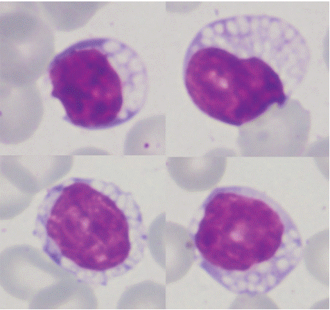
Figure 1: Peripheral blood smear shows many vacuolated lymphocytes (Giemsa stain, magnification x 1000).
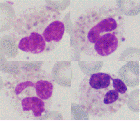
Figure 2: Peripheral blood smear shows abnormal eosinophils (Giemsa stain, magnification x1000).
Enzyme analysis of leukocytes revealed a decrease of Β-D-galactosidase activity and confirmed the diagnosis of GM1 – gangliosidosis, type 1.
Microscopic assessment of a peripheral blood smear in the laboratory provides important information for identifying potentially useful additional tests, understanding the patient's disease, its course and prognosis.
GM1 gangliosidosis is a disease belonging to the group of lysosomal accumulation diseases, caused by systemic accumulation of GM1 ganglioside and its metabolites in cells. Due to its extremely rare incidence and symptoms that overlap with those of other neurodegenerative diseases, diagnosing GM1 gangliosidosis is often a difficult task [4].
Author Statements
Conflict of Interest
The authors declare no conflict of interest.
Funding
The study was performed without external funding.
References
- Chevalier C, Detry G. Vacuolated lymphocytes and abnormal eosinophils in GM1 gangliosidosis, type 1. Br J Haematol. 2012; 156: 293.
- Gitzelmann R, Spycher MA, Adank S, Baerlocher K, Steinmann B. Anomalous eosinophil granulocytes in blood and bone marrow: a diagnostic marker for infantile GM1-gangliosidosis? Eur J Pediatr. 1985; 144: 82-4.
- Salama R, Zhou J. Vacuolated lymphocytes signifying a metabolic disorder in an infant with developmental delay. Clin Case Rep. 2015; 4: 99-100.
- Lee JS, Choi JM, Lee M, Kim SY, Lee S, Lim BC, et al. Diagnostic challenge for the rare lysosomal storage disease: Late infantile GM1 gangliosidosis. Brain Dev. 2018; 40: 383-390.