
Research Article
Austin J Clin Ophthalmol. 2024; 11(2): 1181.
The Urgent Penetrating Keratoplasty by Traumatic Corneal Perforations of a New Trephine Trepanation Technique
Shamurat Amansakhatov¹; Merdan Shakuliev¹; Larisa Ssetinina¹; Mayagozel Zhutdieva²*
¹International Center of Eye Diseases, Ashgabat, Turkmenistan
²International Center of Endocrinology and Surgery, Ashgabat, Turkmenistan
*Corresponding author: Mayagozel Zhutdieva International Center of Endocrinology and Surgery, Ashgabat, Turkmenistan. Email: dr.zhutdieva@mail.ru
Received: January 18, 2024 Accepted: March 08, 2024 Published: March 15, 2024
Abstract
Purpose: Urgent penetrating keratoplasty by traumatic corneal perforations is for try to restore the anatomical integrity of the globe, as well it is to save a vision as much as possible.
Methods: A retrospective review of patients with open corneal injuries was performed in 48 patients (48 eyes) from January 1, 2009 to December 31, 2013 at the International Center of Eye Diseases, Ashgabat, Turkmenistan which carried out various types of surgical techniques with the use of a new trepan. Each patient are depending of trephination technique’s. These patients divided into the following groups: Group I – urgent penetrating keratoplasty with complete removal of the affected area of the cornea, Group II – urgent penetrating keratoplasty with removal of only the central part of the cornea, Group III – urgent penetrating keratoplasty with reconstruction of the eye’s anterior segment or “triple procedure” through by perforated soft contact lens.
Results: In group I, condition of transplant was on the top among 19 patients. Graft transparency have been observed in 16, semi-transparency among 1 patient and hazy among 2. In group II, among 16 patients, clear graft was seen in 9, irreversible in 5 and opacification in 2 patients. The worst results marked in group III, where 5 patients among 13 patients had clear transplant ant, other 3 had semi-transparency, another 5 patients were not clear. (failed test) Optical results were correspondent to biological ones: the best results reported in I-group among 19 patients with visual acuity up to 0.4–0.7 in 7 patients, in the II-group among 16 patients the highest vision to 0.4–0.7 observed in 1 patients and III-group among 13 patients – 0.4–0.7 also 1 patient.
Conclusion: Studies have showed efficiency of trepanation technique with use of a new trepan for penetrating keratoplasty by traumatic corneal perforations. The effectiveness of the operation depends on the preoperational condition of the eye (the shape and size of the wound, time elapsed after injury).
Keywords traumatic corneal perforations, urgent penetrating keratoplasty, trepanation technique, new trephine
Introduction
Worldwide, ocular trauma is an important public health problem which is preventable by its etiology and severity. Outcome depends on factors in the changing environment [1]. It is estimated that, there are approximately 1.6 million people blind from eye injuries, 2.3 million bilaterally visually impaired and 19 million with unilateral visual loss; these facts make ocular trauma the most common cause of unilateral blindness [2]. The incidence rates of ocular trauma requiring hospitalization are reported to be 8.1 per 100 000 persons per year in Scotland [3], 12.6 per 100 000 persons per year in Singapore [4], 13.2 per 100 000 persons per year in the United States [5], and 15.2 per 100 000 persons per year in Australia [6]. Traumatic corneal perforations belong to serious ophthalmic emergencies, which is leading to permanent visual and eyeball loss when it is not promptly treated. Open globe injuries are injuries of the cornea and/or sclera breached and there is a full-thickness wound of the eye ball [7]. Open globe eye injury due to laceration of the cornea or sclera by sharp objects or blunt trauma (contusion) with rupture of the eye possible complications of the crystalline lens, vitreous/retina, and/or optic nerve with the development endophthalmitis. According to some authors the risk of endophthalmitis does not significantly increase in the first 24 or 36 hours [8, 9]. It should be noted that the result of significant structural changes in damaged tissues are often the results of secondary reconstructive operations performed in a long time are unsatisfactory.
The surgical procedures were classified as primary (performed following first presentation) and secondary (performed later following primary procedure). Usually, corneal lacerations are primarily closed with non-absorbable suture such as 10-0 nylon, since the principle of function by known (regular, familiar, famous) trephines does not provide urgent penetrating keratoplasty. From corneal open injuries have observed severe ocular hypotonia and deformation of the damaged cornea results of this state in our clinic have designed/ created new trephine for surgical intervention (surgical intervention by this state in our clinic designed new trephine). The use this trephine there is a possibility circular recipient trephination by hypotonus globes without risk of inner structure damage of eye.
Patients and Methods
This study was a retrospective review of 48 patient’s (48 eyes) surgical treatment with open globe injuries and corneal laceration, which were operated from January 1, 2009 to December 31, 2013, at the International Center of Eye Diseases, Ashgabat, Turkmenistan within carried out various sorts of new trephine surgical techniques which designed in our center. At the presentation of patient’s evaluation have included detailed history of injury, Snellen’s visual acuity, slit lamp examination, intraocular pressure, adequate fundus visualization (by possibility), and ultrasonography (in necessarily). Patients who diagnosed with corneal open injuries as well underwent complete ocular examination were treated by using one of three surgical techniques.
Trephine Design
Trephine consists of two parts: the cutting crown (a-i) and the piston (a-ii) (Figure 1). A significant difference in trepan concerns its piston, which has the shape of a cylindrical rod and moves freely in the vertical direction in the cutting crown. At the distal end there is a flat disk 0.1 mm smaller than the diameter of the cutting crown. The free surface of the disk is a flat rounded plate (Figure 1 b-i rot arrow) and the reverse side has a spherical shape band replicating of corneal curvature (Figure 1 b-ii blue arrow). The flat disk is connected to piston by means of an eccentrically located 1.5-2.0 mm diameter rod. (Figure 1 c-i). The proximal end of the piston as a handle (Figure 1 d-i). For diameters of 5.0–8.0 mm, any type of corneal trephine can be used.
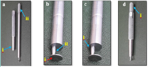
Figure 1: The constituent parts of the trephine.
Surgical Technique
Group I - 19 Patients (First Variant)
This technique is used for corneal traumatic perforations up to 7 mm. By general anesthesia under the operating microscope, the necrotic tissue was removed by forceps and scissors and cleaned of fibrin and fallen damaged tissue (Figure 2a). Holding the trepan in a vertical position above the eye, the disc-shaped end of it was lowered and, in this position, the flat disc of the piston was carefully inserted into the anterior chamber through the corneal wound (Figure 2b). The diameter of the trepan was selected according to the maximum length of the wound. The eccentric arrangement of the connecting rod helped to improve the view and the smooth insertion of the flat disk into the anterior chamber. After complete insertion of the disc-shaped end of the piston into the anterior chamber, it was placed in the central position and the cutting crown was carefully lowered until the latter came into contact with the surface of the (Figure 2c). In this case, the damaged cornea is compressed between the flat disk on the front side of the camera and the cutting crown on top. In this position, they began to rotate the cutting crown, holding the trepan by the handle (Figure 2d). During trepanation, it is important to keep the handle stationary, since its rotation can change the original position of the cutting crown. The donor button was transferred to the recipient bed and fixed in place with four 10.0- monofilament nylon interrupted (at 12, 15,18 and 21 o'clock positions) (Figure 2e), continuous running sutures (Figure 2f). At the end of the operation for air injection to restore anterior chamber. In all cases were injected of gentamycin. All cases were injected of gentamycin.
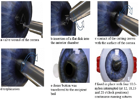
Figure 2: Surgical steps of urgent penetrating keratoplasty with complete removal of the affected area of the cornea (Group I).
Group II – 16 Patients (Second Variant)
This option is already in use for corneal traumatic perforations more than 7 mm. Surgical procedures performed under general anesthesia. Following the anesthesia performed a Barrique eyelid speculum. It is gently inserted to make sure that exert pressure is on the globe. As well making sure not to exert pressure on the globe. Inflammatory membranes over the iris and pupil were mechanically removed by forceps. The reason of large deformation of cornea applied to the peripheral areas after wound fixed by interrupted sutures (Figure 3 a, b) to improve incision’s conditions with trephine the central part of the cornea (Figure 3c). The edges and base of the corneal perforation cleaned with Vannas scissors to remove all damaged inner tissue. The similarly to the first variant, the damaged cornea was excised. In the presence of stellate wounds, separate interrupted sutures were first applied to the edges of the wound directly at the trepanation hole. The trepanation hole form is a regular circle. The corneal donor button was transferred to the recipient bed (Figure 3d) and fixed with 4 interrupted 10.0 Nylon (Figure 3e) sutures and continuous running sutures (Figure 3f). For air injection to restore anterior chamber and a subconjunctival injection of gentamycin.
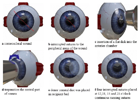
Figure 3: Surgical steps of urgent penetrating keratoplasty with removal of only the central part of the cornea (Group II).
Group III 13 Patients (Third Variant)
Anesthesia is same as in first two groups. The latter option is used for damage the inner structures of the eye (iris, lens) and a risk of extrusion of intraocular contents. The edges of valve wound and surface of iris were mechanically removed with forceps (Figure 4a). Before the trephination of cornea’s central part and excision damaged soft tissue (Figure 4 a, b) and soft contact lens 0 diopters, 14.0-15.0mm with paracentral hole ø 5 mm prepared in advance and placed on its surface (Figure 4c).
A lensectomy with IOL implantation or "triple procedure" was performed through a paracentral hole in a soft contact lens, turning in the right direction. Donor corneal disc was placed in recipient bed and fixed first 4 knoten sutures nylon 10.0 and continuous running sutures (Figure 4d, e, f). The anterior chamber to restore for air injection. At the conclusion of surgery gentamicin was injected subconjunctival.
Results
Epidemiological Findings
From 48 patients, 35 patients (73.0%) were male and 13 (27.0%) were female (Figure 5). Divided by age groups, on the one hand in the group between 21-50 years there was increased occurrence and on the other hand in the group from 51 years. The main age was (32.3 ± 5.9) years (min. 1, max. 60 Years) (Figure 6). The right eye was more often affected (66.7%) compare to the left eye (33.3%).
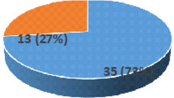
Figure 5: Demographic profile of our study group. Sex disribution patients with traumatic corneal perforations.
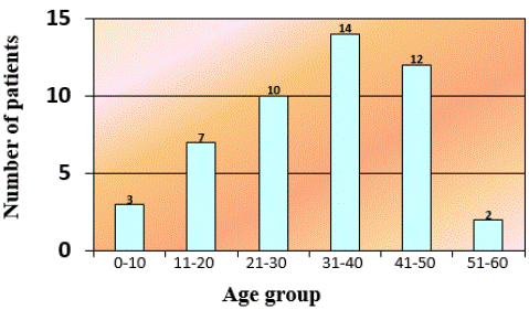
Figure 6: Age disribution patients with traumatic corneal perforations.
In this study among the patients with open corneal injuries the prevalence of men, namely male 41/85.5%, female 7/14.5% range 10–60 years, peaks of incidence were recorded at the age of 18 to 30 years.
Table 1 gives an overview of the documented causes of accidents and the number of penetrating corneal injuries.
    Cause
    Incidence
    Percent %


    Road accident
    12
    (25%)


    Tree/branch
    7
    (14,6%)


    Stone
    5
    (10,4%)


    Garden tools
    5
    (10,4%)


    Metallic objects
    4
    (8,3%)


    Toy
    4
    (8,3%)


    Punch
    3
    (6,2%)


    Falling from a height
    2
    (4,2%)


    Expander
    2
    (4,2%)


    Injuries caused by animals
    1
    (2,1%)


    Piece of    wood
    1
    (2,1%)


    Bottle cap
    1
    (2,1%)


    Door handle
    1
    (2,1%)


    Total
    48
    (100%)
Table 1: Traumatic agents in 48 patients with penetrating corneal perforations.
Graft Clarity after Urgent Penetrating Keratoplasty
The status of transplant at after urgent penetrating keratoplasty are summarized in Table 2 and clinical photographs in Figure 7. Graft rejection in the first group clear graft 16/84.2%, reversible 1/5.2%, irreversible 2/10.6%. In the second group graft transparency was better than in the third group exactly: clear 10/62.5%, semi-transparency 2/12.5%, hazy 4/25%. Graft survival was poor for cases in third group: clear 2/15.3%, semi- transparency 7/54%, hazy 4/30.7%

Figure 7: The clear graft rejection of transplantat after urgent penetrating keratoplasty.
    The status    of transplantat
    Number of cases/Percentage


    clear graft
    versible
    irreversible


    Group I
    16
    1
    2
    19 (39.6%)


    Group II
    10
    2
    4
    16 (33.4%)


    Group III
    2
    7
    4
    13 (27.0%)
Table 2: The graft rejection after urgent penetrating keratoplasty with different surgical techniques (Groups I, II, III).
    Visual acuity
    Number of cases /Percentage


    0.01–0.04
    0.05-0.09
    0.1-0.3
    0.4-0.7


    Group I
    2
    2
    8
    7
    19 (39.6%)


    Group II
    6
    5
    4
    1
    16 (33.4%)


    Group III
    5
    4
    3
    1
    13 (27.0%)
Table 3: Optical results of 48 patients after urgent penetrating keratoplasty with different surgical techniques.
Vision Outcome
In our study the preoperative visual acuity in all patients ranged from light perception to 0.05. Visual acuity after urgent penetrating keratoplasty shown in Table 2. The table shows that in the first group, 15 operated patients had visual acuity of 0.1 or higher, which indicates the effectiveness of this operation. In the second group, 5 patients had visual acuity in the range of 0.1-0.7, and 11 patients had visual acuity in the range of 0.01-0.09. Low vision was primarily observed in 6 patients with semi-transparency and hazy grafts. In addition, the same patients had destruction of the vitreous body and secondary cataract also the formation of a fibrous film in the pupil area pupillary membrane. The results of the observation showed that the severity of the injury, especially damage to the lens, was essential for the outcome of urgent keratoplasty in the second option, so the biological and optical efficiency of the operation was significantly inferior to the first option. In the third group, the optical results are significantly inferior to the first group.
Discussion
Penetrating eye injuries are one of the important causes of severe visual impairment and is an ophthalmic emergency that needs immediate treatment. To close the non-traumatic defect of the cornea there is an arsenal of materials both biologic: conjunctival flap [10], conjunctivoplasty/or multilayered amniotic membrane transplantation [11,12], fibrin glue [13] and synthetic derivatives: cyanoacrylate glue [14,15], multilayered Gore-Tex patches [16,17]. All available methods have been shown to be effective in the closure of small corneal perforations (up to 3mm in diameter). The choice of the treatment depends on the size and location of the perforation and status of underlying diseases.
Our date is consistent with those of the authors indicating that men are at significantly higher risk (approximately four times higher) than women. Almost half of all reported injuries occur in people aged 18- 45 years. [18-20].
Though the prognosis of open globe injuries has improved with the development of advanced microsurgical techniques and better understanding of tissue reaction to trauma [21]. A review of experience indicates a very significant impact of eye injuries in terms of medical care, needs for vocational rehabilitation and great socioeconomic costs [2].
Generally, if the perforation is very small (diameter=1 mm), a temporary cyanoacrylate glue application can be used Refojo et al. [22]. Over 20 years ago, Bessant and Dart [23] described a technique for trephination involving the use of cyanoacrylate glue and a lamellar graft in 10 eyes with peripheral inflammatory corneal ulcers that were either perforated or pre-perforated. In these cases, there was a small perforation in the middle of an extensive ulcer, making it possible to seal the hole with just a few drops of cyanoacrylate glue. The anterior chamber was restored by injecting a viscoelastic solution, allowing lamellar dissection. This technique is very useful, but it can only apply to perforations with a very small diameter.
For slightly larger perforations, cyanoacrylate glue alone is not sufficient. Por et al. [24] reported three cases of perforations of 2–4mm in diameter, there they proposed a fibrin glue injection into the anterior chamber and once the cyanoacrylate glue was correctly centered on the perforation. This double seal restored the anterior chamber allowing lamellar dissection. Nevertheless, fibrin glue injection requires a minimum degree of sealing with cyanoacrylate glue, which is difficult to achieve when the perforation is more than 2mm in diameter.
All described manipulations refer to non-traumatic corneal perforations. However, due to the increase in the number of severe penetrating wounds, primary reconstructive operations performed at the time of surgical treatment of the wound began to occupy a special place. The difficulty of performing such operations is that several manipulations are performed simultaneously on different tissue of the eye such as urgent penetrating keratoplasty, iridopathy and simultaneously with cataract extraction and IOL implantation. The technique of trepanation with new trephine for urgent penetrating keratoplasty in satisfactory conditions on patients with a corneal perforation of more than 4mm in diameter.
First variation is surgeries are offered for stellate and linear penetrating wounds of the cornea, including its entire thickness and damage with a diameter of up to 7 mm, but with fairly smooth edges.
Second variation is indicated in cases where there is a wound with a diameter of more than 7 mm. since the dilution of the wound is associated with additional trauma, it is recommended to first apply stitches to the edges of the wound in order to create a circle and an opportunity for trepanation. Third variation is recommended for smashed wounds with damage to the inner membranes of the eye, as well as in the presence of a corneal defect. However, due to severe hypotension, through trepanation is not performed, but only the border of the future incision is marked, followed by excision of intended corneal area with a blade and scissors and replacement with a donor cornea [25].
In recent years, we have refused to them the Fleering ring, which creates a number of inconveniences during the operation. During urgent keratoplasty, we use a soft contact lens with a diameter of 14-15 mm with a paracentral hole with a diameter of 5 mm, which is very convenient to use. When using a soft contact lens, better working conditions are created by reducing the open surface of the wound in total and subtotal keratoplasty, which can be rotated in the desired direction, which means that the risk prolapse of iris, lens and corpus vitreum is significantly reduced.
Climatic conditions of Turkmenistan characterized by a sharply continental climate with a predominance of a long hot period, with dry and hot weather, in turn, also contributes to an increase in infection and has a negative impact on the course of the post-traumatic period [26].
The following factors were among the main factors affecting visual functions and the nature of graft healing: the size, shape and location of the wound, as well as the degree of damage to the inner structures of the eye and the age of the eye injury. The frequency of cataract formation after severe corneal injuries, the need for late surgical treatment using urgent keratoplasty - all this made it necessary to develop a choice of the most rational methods of primary treatment of severe corneal injuries.
In conclusion, this study suggests that the severity of injury, time of presentation and institution of prompt treatment are also very important determinants in the final visual and anatomical outcome. Despite the great possibilities of microsurgery, achieving complete rehabilitation of patients after primary surgical treatment is a difficult task. Often, the cause of vision loss or death of the eye is not the injury itself, but the complications that occurred after it. Therefore, modern urgent assistance should be comprehensive as much as possible. The proposed technique of trepanation using a new trepan is an alternative method of surgery for open corneal injuries.
Author Statements
Acknowledgement
The authors would like to thank Dr. med. Merjen Allaberdyeva, International Center of Neurology, Ashgabat, Turkmenistan, for linguistic revision.
Competing Interests
The authors declare that they have no competing interests.
References
- Wong TY Epidemiological characteristics of ocular trauma in the US Army. Arch Ophthalmol. 2002; 120: 1236.
- Negrel AD, Thylefors B. The global impact of eye injuries. Ophthalmic Epidemiol. 1998; 5: 143–169.
- Desai P, MacEwen CJ, Baines P, Minassian DC. Incidence of cases of ocular trauma admitted to hospital and incidence of blinding outcome. Br J Ophthalmol. 1996; 80: 592–596.
- Wong TY, Tielsch JM. A population-based study on the incidence of severe ocular trauma in Singapore. Am J Ophthalmol. 1999; 128: 345–351
- Klopfer J, Tielsch JM, Vitale S, See LC, Canner JK. Ocular trauma in the United States: eye injuries resulting in hospitalization, 1984 through 1987. Arch Ophthalmol. 1992; 110: 838–842.
- Fong LP. Eye injuries in Victoria, Australia. Med J Aust. 1995; 162: 64–68.
- Kuhn F, Morris R, Mester V, Witherspoon CD. Ocular Traumatology. Springer, 1.1 Terminology of mechanical injuries: the Birmingham Eye Trauma Terminology (BETT). 2008; 3-11.
- Thompson WS, Rubsamen PE, Flynn Jr HW, Schiffman J, Cousins CW. Endophthalmitis after penetrating trauma. Ophthalmology. 1995; 102:1696-701.
- Barr C. Prognostic factors in corneoscleral lacerations. Arch Ophthalmol. 1983; 101: 919-924.
- Geria RC, Zarate J, Geria MA. Penetrating keratoplasty in eyes treated with conjunctival flaps. Cornea 2001; 20: 345–349.
- Hanada K, Shimazaki J, Shimmura S, Tsubota. K Multilayered amniotic membrane transplantation for severe ulceration of the cornea and sclera. Am J Ophthalmol. 2001; 131: 324–331.
- Lee SH, Tseng SC. Amniotic membrane transplantation for persistent epithelial defects with ulceration. Am J Ophthalmol. 1997; 123: 303–312.
- Moschos M, Droutsas D, Boussalis P, Tsioulias G. Clinical experience with cyanoacrylate tissue adhesiv?. Doc Ophthalmol .1996; 93: 237-245.
- Lagoutte FM, Gauthier L, Comte PR. A fibrin sealant for perforated and preperforated corneal ulcers. Br J Ophthalmol. 1989; 73: 757–761.
- Rüfer F, Eisenack J, Klettner A, Zeuner R, Hillenkamp J, Westphal G, et al. Multilayered Gore-Tex patch for temporary coverage of deep noninfectious corneal defects: surgical procedure and clinical experience. Am J Ophthalmol. 2011; 151: 703–713.
- Amm M, Nölle B. Gore-Tex-patch-Aufnähung bei immunologisch bedingten Hornhautulzeration. Klin Monatsbl Augenheilkd. 2002; 21: 735-739.
- Gauthier L, Lagoutte F. Use of a fibrin glue (Tissucol) for treating perforated or pre-perforated corneal ulcer. J Fr Ophtalmol. 1989; 12: 469–476.
- Thylefors B. Epidemiological patterns of ocular trauma. Aust NZ J Ophthalmol. 1992; 20: 95-98.
- Schein OD, Hibberd PL, Shingleton BJ, Kunzweiler T, Frambach DA, Seddon JM, Fontan NL, Vinger PF. The spectrum and burden of ocular injury. Ophthalmology. 1988; 95: 300-305.
- Mac Ewen CJ. Eye injuries: a prospective survey of 5671 cases. Br J Ophthalmology. 1989; 73: 888-894
- Mac Ewen CJ. Ocular injuries. J R Coll Surg Edinb. 1999; 44; 317-323.
- Refojo MF, Dohlman CH, Ahmad B. Carroll JM, Allen JC. Evaluation of adhesives for corneal surgery. Arch Ophthalmol. 1968; 80: 645–656.
- Bessant DA, Dart JK. Lamellar keratoplasty in the management of inflammatory corneal ulceration and perforation. Eye London. 1994; 8: 22–28.
- Por YM, Tan YL, Mehta JS, Tan DTH. Intracameral fibrin tissue sealant as an adjunct in tectonic lamellar keratoplasty for large corneal perforations. Cornea. 2009; 28: 451–455.
- Amansakhatov Sh. Indications and efficiency of keratoplasty in inflammatory processes of cornea in Turkmenistan. Thesis for a candidate of medical science degree. Odessa. 1981; 127-129.
- Sereteli EK. The seasonality of conjunctival flora of normal human eye in conditions of Turkmenistan. Abstract book of scientific-clinical center of eye diseases named S. Karanov. Ashgabat. 1971; 59-61.