Abstract
Purpose: The prevalence of ocular involvement in patients with systemic sarcoidosis ranges widely from 13% to 79%. Choroidal granuloma is a relatively rare lesion that may mimic a choroidal tumor in the fundus, making diagnosis and treatment difficult.
In this study, we report the case of a solitary sarcoid choroidal granuloma.
Case Report: A 26 year-old female patient presented to the ophtalmology emergency departement with the primary complaint of decreased visual acuity in her left eye. Upon examination, the visual acuity in the concerned eye was light perception. The fundus evaluation revealed a subretinal tumor mass with extentive exudative retinal detachement in her left eye. Ultrasound B-scan examination showed a localized choroidal elevated mass lesion over the posterior pole with high surface and internal reflectivity. MRI scan revealed elevated enhancing choroidal lesion temporal to the left optic disc. No etiology could be found and a potiental malignancy couldn’t be ruled out. The patient underwent enucleation of the left eye. The histopathological exam findings were concording with the diagnosis of a sarcoidosis granuloma.
Conclusion: Accurate diagnosis of solitary choroidal granuloma as a manifestation of sarcoidosis remains difficult. Differentiating these lesions from amelanotic melanoma and metastatic tumors of choroid needs thorough investigation.
Keywords: Sarcoidosis; Choroid; Granuloma
Introduction
Sarcoidosis is a chronic multisystemic granulomatous disorder characterized by the accumulation of non- caseating granulomas in the involved tissues. The prevalence of ocular involvement in patients with systemic sarcoidosis ranges widely from 13% to 79%. Ocular involvement is the presenting symptom in 20-30%. Ophthalmic manifestations can be isolated or associated with other organ involvement. Ocular sarcoidosis can involve any part of the eye and its adnexal tissues and may cause uveitis, episcleritis/scleritis, conjunctival granuloma, optic neuropathy, lacrimal gland enlargement, and orbital inflammation. Choroidal granuloma is a relatively rare lesion that may mimic a choroidal tumor in the fundus, making diagnosis and treatment difficult. In this study, we report the case of a solitary sarcoid choroidal granuloma revealing sarcoidosis.
Case Report
A 26-year old female patient presented to the ophtalmology emergency department with the primary complaint of painless decreased visual acuity of 2 months in her left eye. Her medical and family history was unremarkable.
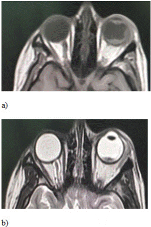
Upon examination, the visual, best-corrected visual acuity was 10/10 in the right eye, and reduced to light perception in the left eye. Slit-lamp examination in both eyes revealed quiet anterior chamber with no cells flare or synechiae. Intraocular pressure was 13 mmHg in both eyes. Gonioscopy showed no angle abnormalities in both eyes. Fundus examination with indirect ophthalmoscopy and slit-lamp biomicroscopy showed an elevated mass in the left eye, massive subretinal fluid surrounding it and total retinal detachment. There was no abnormal pigmentation or abnormal vessels associated with the mass. There were no exudates over the pars plana (Figure 1). The fundus examination of the right eye was normal. Ultrasound B-scan examination of the left eye revealed a diffuse retinochoroidal elevated mass lesion over the posterior pole with high surface reflectivity and low internal reflectivity. Extensive subretinal fluid was detected surrounding the lesion. The lesion measured 4.5 mm in height, 20 mm in radial diameter, and 4.4 mm in circumferential diameter. There was no T sign, choroidal excavation or orbital shadowing. Magnetic Resonance Imaging (MRI) showed a mass infiltrating the head of the optic nerve, hypointense on T1 and T2-weighted images and enhanced after injection of gadolinium.

Figure 1: Retinophotoghraphies of the left eye showing the extensive exudative retinal detachment with the underlying mass.

Figure 2: MRI scans showing the choroidal mass: a) T1 weighed Scan; b) T2 weighed MRI Scan.
Systemic investigations including blood cell count, erythrocyte sedimentation rate, serum angiotensin converting enzyme, Quantiferon TB-Gold, tuberculin test, toxoplasmosis serology, syphilis serology and tumor markers were within the normal range. The chest CT-scan was normal. Positron Emission Tomography (PET) scan done in order to rule out any systemic malignancy and showed no abnormalities.
Despite the investigations, no etiology could be found and a potential malignancy could not be ruled out. The visual acuity decreased to no light perception. The patient underwent enucleation of the left eye. The histopathological exam findings were concording with the diagnosis of a sarcoidosis granuloma. At 2 years follow up, the patient had not shown any other manifestations of ocular or systemic sarcoidosis.
Discussion
Sarcoidosis is a multisystem disorder, consisting of granulomatous inflammation of unknown etiology with a histological hallmark of noncaseating granulomas composed of epithelioid cells and Langerhans giant cells. Posterior segment findings in ocular sarcoidosis have been shown to occur in 14–28% of patients, including vitritis, retinal vasculitis, chorioretinitis, vascular occlusions, retinal neovascularization, and granulomas involving retina, optic nerve, and choroid [1].
Choroidal granulomas manifesting as the sole lesion in ocular sarcoidosis has been previously described. The largest case series of choroidal granulomas concerned nine patients with biopsy-proven sarcoidosis. Eight patients presented with solitary choroidal granulomas, and one patient presented with multifocal involvement [1]. Reportedly, there is no sex difference and it most commonly occurs in individuals 60 years of age or less.
Unlike usual ocular sarcoidosis, this pathology commonly shows mild inflammation in the anterior chamber or the vitreous body and often presents diagnostic difficulties. The lesion is well-defined and pale yellow, and the size ranges between 1 and 10 PD. Fluorescein angiography examination shows low fluorescence or punctate hyperfluorescence corresponding to the lesion at the early phase and hyperfluorescence from the middle to late phase [2]. It has been reported that indocyanine green angiography examination can show low fluorescence until the late phase, and that OCT findings show a homogenous shadow under the retina [2]. Fundus findings of this disorder mimic those of a choroidal tumor in many respects, and thus, a general examination including head CT/magnetic resonance imaging is often needed.
Lesions are hypointense with respect to orbital fat on T1- and T2-weighted images and enhance strongly with contrast administration [3]. The differential diagnosis is that of an orbital mass, namely, orbital lymphoma, metastases, Weger granuloma, or orbital pseudotumour.
The concensus conference of the International Workshopon Ocular Sarcoidosis [4] identified seven signs in the diagnosis of intraocular sarcoidosis: mutton-fat keratic precipitates /small granulomatous KPs and/or iris nodules (Koeppe/Busacca), trabecular meshwork nodules and/or tent-shaped peripheral anterior synechiae, vitreous opacities displaying snowballs/strings of pearls, multiple chorioretinal peripheral lesions (active and/or atrophic), nodular and/or segmental peri-phlebitis (+/- candlewax drippings) and/or retinal macroaneurism in an inflamed eye, optic disc nodule(s)/granuloma(s) and/or solitary choroidal nodule, and bilaterality. The laboratory investigations or investigational procedures that were judged to provide value in the diagnosis of ocular sarcoidosis in patients having the above intraocular signs included : negative tuberculin skin test in a BCG- vaccinated patient or in a patient having had a positive tuberculin skin test previously, elevated serum Angiotensin Converting Enzyme (ACE) levels and/or elevated serum lysozyme, chest x-ray revealing Bilateral Hilar Lymphadenopathy (BHL), abnormal liver enzyme tests, and chest CT scan in patients with a negative chest x-ray result. For our patient, all the investigations were negative.
With respect to ocular sarcoidosis, previous reports described the use of steroid pulse therapy for panuveitis or optic neuropathy yet the publiched reports on choroidal granuloma are scarce. However, a case of a refractory patient with a large choroidal granuloma, panuveitis, and optic disc edema being treated with high-dose intravenous steroid therapy was reported [5]. A choroidal granuloma associated with sarcoidosis often responds well to oral steroid therapy, with the therapy often being started with a dose equivalent to prednisolone 20–80 mg, and then gradually tapered and discontinued in 3–10 months [6]. Some previous studies reported that injection of steroid into the Tenon capsule or the vitreous body was effective [7].
Conclusion
In conclusion, accurate diagnosis of solitary choroidal granuloma as a manifestation of sarcoidosid remains difficult. Differentiating these lesions from amelanotic melanoma and metastatic tumors of choroid needs thorough investigation.
References
- Kawaguchi T, Hanada A, Horie S, Sugamoto Y, Sugita S, Mochizuki M. Evaluation of characteristic ocular signs and systemic investigations in ocular sarcoidosis patients. Jpn J Ophthalmol. 2007; 51: 121-6.
- Modi YS, Epstein A, Bhaleeya S, Harbour JW, Albini T. Multimodal imaging of sarcoid choroidal granulomas. J Ophthal Inflam Infect. 2013; 3: 58.
- Mysore N, Gonçalves FG, Chankowsky J, del Carpio-O’Donovan R. Adult orbital masses: A pictorial review. Can Assoc Radiol J févr. 2012; 63: 39-46.
- Mochizuki M, Smith JR, Takase H, Kaburaki T, Acharya NR, Rao NA, et al. Revised criteria of International Workshop on Ocular Sarcoidosis (IWOS) for the diagnosis of ocular sarcoidosis. Br J Ophthalmol. 2019; 103: 1418-22.
- Kobayashi T, Takai N, Sato T, Tada R, Maruyama E, Shouda H, et al. A case of large sarcoid choroidal granuloma treated with steroid pulse therapy. Case Rep Ophthalmol. 2020; 11: 112-9.
- Verma A, Biswas J. Choroidal granuloma as an initial manifestation of systemic sarcoidosis. Int Ophthalmol. 2010; 30: 603-6.
- Chan WM, Lim E, Liu DTL, Law RWK, Lam DSC. Intravitreal triamcinolone acetonide for choroidal granuloma in sarcoidosis. Am J Ophthalmol. 2005; 139: 1116-8.
